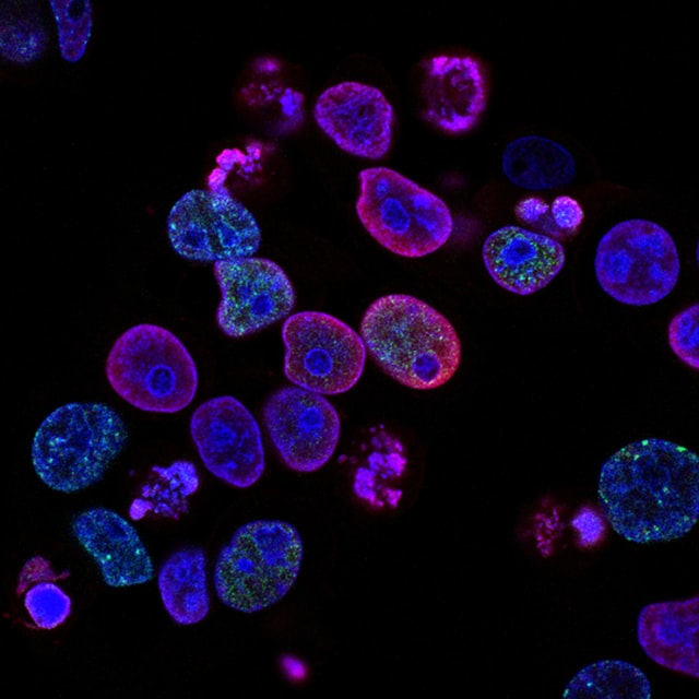
The fate of a cell refers to the description of future of the cell. In normal way we can say that fate of a cell means, it describes what cell become in due course of development. Therefore fate map of a cell can be defined by labelling and observing the cell’s division, differentation, and migration in it’s normal developmental process through a diagram. In fate map we can depicts that which part of the egg or early embryo determine to form which specific tissue or structure at later stage or advanced stage of development.
Another benifit of constructing fate map is to understands the lineage and migration of progenitor cells, through advanced techniques which is also helps to discover gene regulatory networks and signalling pathways.
Construction of fate maps :
Fate map is a experimental tool to deduce reorganization of cells from early stage of development to later or advanced stage of development. In this process of construction labelling of a cell at a recorded position marked by natural colour or artificial dye and observing the same cell or it’s progeny cells position at later stage. Various techniques have been instructed for construction of fate map, among which tracing through natural colour and artificial markings are very much useful.
Natural Markings :
Fate mapping can be done through natural marking or pigments. In cytoplasm of certain organism contain natural pigments,which can be helpful for fate mapping. Such example is the pigments present in cytoplasm of egg of certain ascidian. The fertilized egg contain four colored centres i.e. an upper hemisphere of light protoplasm, an yellow crescent posterio-ventrally, a grey crescent anterio-dorsally and a vegetal area of dark grey yolk substance. The fate of these areas can be acknowledged very easily and it has been seen that the upper clear cytoplasm contains the material for epidermal ectoderm, the grey crescent area get differentiated into the prospective neuroectoderm and notochord, the yellow crescent becomes the prospective mesoderm and the dark yolk area responsible for formation of prospective endoderm.
Artificial Markings :
Artificial markings includes three methods of marking or labelling in early blastomere, which are as follows :-
i) Vital staining :
Vital staining method was first introduced by Waller Vogt in 1928. Vital stains such as methylene blue, neutral red, bismark brown, janus green and Nile blue sulphate, which are life friendly beacuse it doesn’t harm the cell. Due to the persistence or last long character and cell friendly activities of the dye makes it possible to determine the ultimate location of marked area in late embryo. In this method first of all soaking of a piece of agar with vital dye takes place and positioning it at the required location of embryo. After that the stain will diffuse into blastomere and ultimately the cell get stained. Here, in this technique descendants of the marked cells also become stained. Hence, it is easy to mark the differentiated embryo, and the parts stained indicates the fates of the original stained blastomeres. Suppose a cell has stained at early embryo and found in the gut region at later embryo, then it could be say that the early stained cells of blastomere is marked for gut development. In this way different colour can be used and marked at specific location of embryo and accordingly the formation of different organ can be know easily.
ii) Carbon particles marking :
This technique was introduced by Spratt in 1946 in order to know the process of formation of primitive streak in chick. Here, a tiny particle of carbon has to be apply over the surface of blastomere and the carbon particle get stick to the cell surface which enable the investigator to trace the movements of cell and fate of the blastomere can be access easily.
iii) Radioactive isotope labelling :
We well known about the characteristics of radioactive isotopes as they emit radiation like alpha, beta, and gamma rays. Due to this property radioactive isotopes are used in tracer experiments. Isotopes such as C14 and P32 are used to labelled the early blastomere and in due course of development monitored of this isotopes revealed the fate of blastomere.
